Cellular structure of organisms
the human body is made of cells;
cell has features of living beings.
divide cells and organisms into Prokaryotes and Eukaryotes;
recognize structures contained in bacterial, plant and animal cells;
show the importance of cellular structures: cell membrane, cytoplasm, cell nucleus, chloroplasts, mitochondrion, vacuole, cell wall;
show the relationship between cell structure and its function.
Cells form the organism
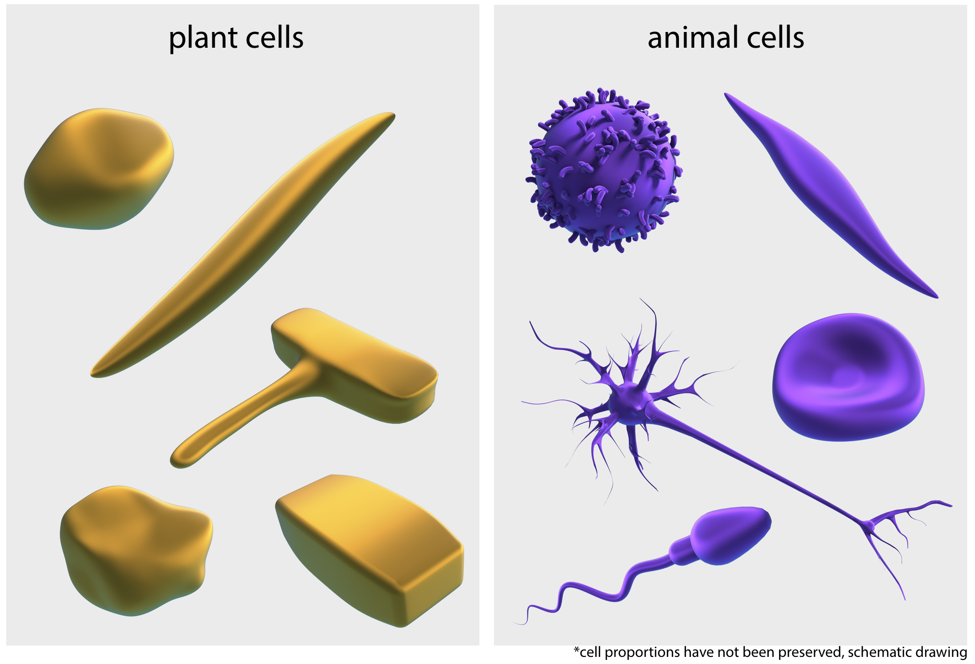
CellsCells are the basic elements that build every organism. Their size and shapes are very diverse, because they depend on the functions fulfilled. The smallest are bacterial cells that have on average one thousandth of a millimeter, or one micrometer (1 μm) in length. The largest cells are found in plants - hemp fiber can reach a length of 50 cm. All cells consist of smaller elements. One of them is the cell nucleusthe cell nucleus - due to its presence or its absence, cells are divided into prokaryotic (bacteria) and eukaryotic (protists, plants, animals,fungi).
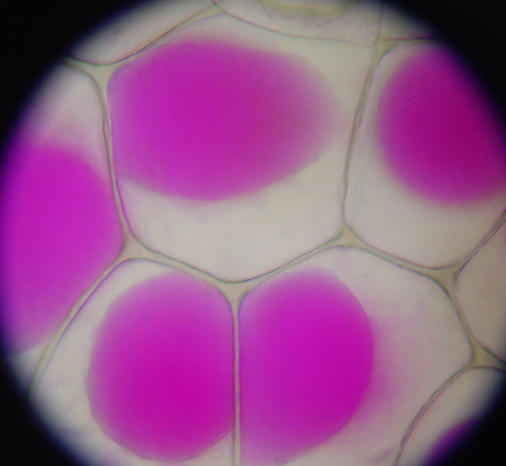
Description of the shape and arrangement of onion leaf epidermal cells.
the epidermis of the onion storage leaf,
microscope slides: basic and covers,
microscope,
dropper,
water,
knife,
preparation needle,
plasticine (modeline).
Divide the onions into scales. From the inside of the scale, remove the needle with a piece of epidermis that looks like a translucent foil. Place the epidermis in a drop of water on the slide.
Cover the prepared preparation with a coverslip so that there are no air bubbles beneath it.
Observe the epidermis at low magnification. Pay attention to the shape and arrangement of the cells.
Make cell models from plasticine. Remember that they are solids. Make a model of epidermis from several plasticine cells.
The arrangement of epidermis cells is related to the function they perform. The spatial shape of the cells can be observed through the microscope, focusing alternately on the upper and lower cell walls.
Spatial organization of the cell
The cells of plants, animals, fungi and bacteria differ in size, shape, internal structure but show a common construction plan. The components of the cells can be divided into living components of a cell, which include the cell membranecell membrane and the cytoplasmcytoplasm together with the structures suspended in it, and non‑living components of a cell, which are the cell wallcell wall and the vacuole.
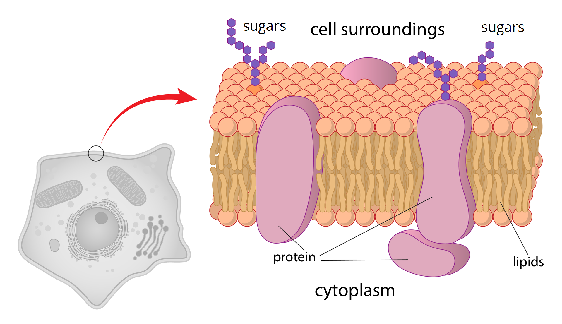
The plasma membrane separates the cell from the environment. It is often called a „cell guardian”, because it is responsible for protecting its interior and for its contact with the environment. In plants, fungi and bacteria, there is a stiff, thick, cell wall outside of the membrane. It can be compared with a box that gives the shape of a cell.
A cytoplasm - a jelly substance composed of water and dissolved in it organic and inorganic compounds, surrounded by a plasma membrane. Cytoplasm is an environment in which chemical reactions necessary for life take place - it is semi‑fluid, and cellular structures are suspended in it. The cytoplasm allows them to move and exchange substances between them. In the cytoplasm, membranes are built like a plasma membrane and form a membranous structures like endoplasmic reticulum or Golgi apparatus.
Checking if the cells exhibit features of life.
Canadian waterweed,
microscope equipment.
An hour up to two hours before the planned observation, take off from the tip of the leaf the leaves of Canadian waterweed and transfer it to water at a temperature of 25‑30 ° C.
Place the leaf in a drop of water on the slide. Cover it with a coverslip so that there are no air bubbles.
Observe the movements of the cytoplasm and the components suspended in it.
Compare cell structures observed under the microscope shown in figure 2. Name them.
Draw an image of the selected cell viewed under the microscope. Describe your drawing and mark the direction of the cytoplasm with the arrows.
The cytoplasm moves in the cell, carrying chloroplastschloroplasts with it. Movement is one of the attributes of life.
Cell structures
The cell membrane is so thin that it cannot be seen in the light microscope. It consists of proteins and fats (lipids). The membrane is semi‑permeable: it permits the penetration of water and other small molecules that through pores (holes), and the larger particles are retained. Their uptake can be done according to the needs of the cell, but it usually requires energy consumption. The membrane receives and conducts stimuli (information) from the environment. It's flexible, therefore it allows cells to change shape.
Study of cell membrane function based on observation of its model.
jar,
distilled water,
glucose,
funnel,
cellulose foil
beaker.
Prepare a 10% glucose solution. In a beaker, measure 450 ml of water and add 50g of sugar, mix until all sugar crystals are dissolved.
Pour distilled water into the jar.
Cover the outlet of the funnel with cellulose foil and pour the glucose solution.
Insert the solution funnel into the jar with distilled water.
Observe the solution level in the funnel. Check the taste of the liquid in the jar and in the funnel.
The film – constituting the cell membrane model – is semipermeable. Water passes through it (from the jar to the funnel), while glucose is stopped.
The cell nucleus can be seen in a light microscope. It is most often a spherical structure surrounded by cytoplasm. The nucleus stores genetic material that contains information about cell structure and functioning. Thanks to this, it controls all its life activities, including division into daughter cells. The nucleus is surrounded by a double nuclear membrane with numerous orifices (pores) allowing the contact of the nucleoplasm with the cytoplasm of the cell. Nucleic acid molecules which including „instructions on the functioning of the cell and whole organism” penetrate from the cell nucleus into the cytoplasm. In the same way, the substances needed there get through from the cytoplasm to the cell nucleus.
MitochondriaMitochondria are invisible in a light microscope. They are surrounded by a double protein‑lipid membrane: the outer one is smooth, the inner one - pleated and forms numerous, lamellate protrusions called cristae. There is a process of cellular respiration on the surface of the inner membrane. It consists in the release of energy accumulated in organic compounds. The more cristae in the mitochondria, the more intense respiration process is.
Mitochondrial matrix (matrix) an amorphous liquid substance that fills the interior of the mitochondrion.

The largest concentrations of mitochondria occur in skeletal muscle cells as well as nerve and secretory cells. Less mitochondria are found in skin cells, for example. Explain why there are many more mitochondria in some tissues than in others.
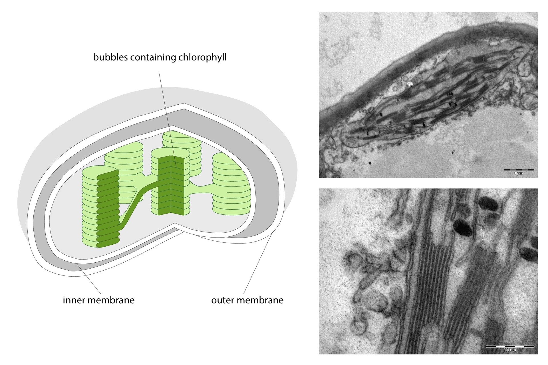
Chloroplasts are well visible in plant cells viewed under a microscope. They contain a green dye - chlorophyll, which has the ability to absorb light energy. The process of photosynthesis takes place in chloroplasts. It consists in the production of organic compounds (sugars) from inorganic compounds (water and carbon dioxide).
The cytoplasm has an irregular plasma membrane system. These are membranes of the endoplasmatic reticulum that divide the cell's interior into smaller spaces and allow simultaneous overlap of various processes and transport of substances between individual cell elements. On the surface of some of the reticular membranes there are lumpy corpuscles - ribosomes. They participate in the synthesis of proteins.
Another membranous structure suspended in the cytoplasm is the Golgi apparatusGolgi apparatus. It is made up of several types of vesicles: large, flattened, arranged parallel to one another and small, spherical vesicles tearing away from them. The Golgi apparatus is involved in the synthesis and modification of some organic compounds, it also collects proteins and other substances.
Lysosomes are small irregularly shaped vesicles surrounded by a single protein‑lipid membrane produced by the Golgi apparatus. They occur in eukaryotic cells. Thanks to the presence of digestive enzymes, they participate, among others in the digestion of nutrients and damaged organelles.
Explain why the onion leaf epidermal cells forming the bulb (storage organ) do not contain chloroplasts, and the leaf epidermal cells of the canadian waterweed are filled with them.
Plant cells as well as fungal and bacterial cells are covered not only by the cell membrane. They have an additionally stiff cell wall. The basic building material of the wall is water‑insoluble sugar - cellulose (or chitin in most fungi). In young cells, the wall is thin and elastic (so as not to limit the growth of cells), but with age it becomes thicker. The cell wall strengthens the protection against the penetration of microorganisms, protects the cell against damage and gives it a shape.
VacuolesVacuoles are mainly found in plant and fungal cells that provide cells with firmness. They are vesicles surrounded by a single, membrane filled with cellular juicecellular juice. Additionally, they contain superfluous substances and secretions collected periodically and used as needed. In animals, vacuoles occur exceptionally only in adipose tissue cells and are used to store its reserves.
There are many vacuoles in young plant cells. As the cells mature, the vacuoles combine to form one central vacuole.
Explain why plants – unlike animals – are not able to move.
Match pairs.
ściana komórkowa, wakuola, dyfuzja, komórka, błona komórkowa, sok komórkowy, cytoplazma, chloroplasty, jądro komórkowe
| cell membrane | |
| chloroplasts | |
| cytoplasm | |
| diffusion | |
| the cell nucleus | |
| cell | |
| vacuole | |
| cellular juice | |
| cell wall |
Summary
Cells – due to the presence or absence of the cell nucleus – we divide into procaryotic and eucaryotic.
Each cell is surrounded by a cell membrane.
In animal cells there are cell structures such as: cell membrane, cytoplasm, cell nucleus, mitochondrium, endoplasmic reticulum, Golgi apparatus.
Plant cell – in contrast to animal – has chloroplasts, cell wall and vacuoles.
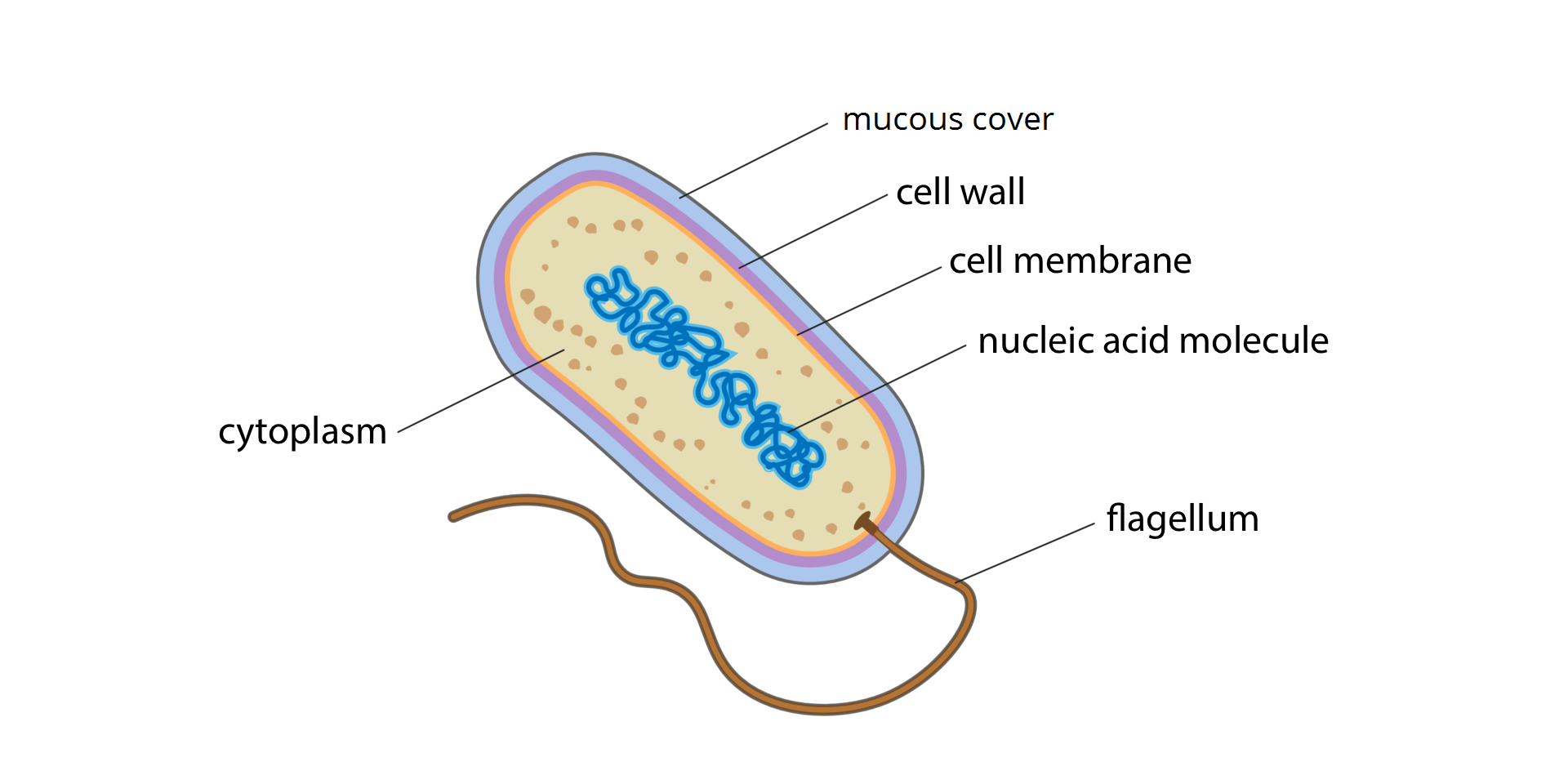
The bacterial cell has no cell nucleus or any membranous structures in cytoplasm.
The cell membrane is semipermeable – it allows the osmotic penetration of water, but retains large particles.
Take a plant cell model from the vial of medicines, food film, beads and other small objects.
List two similarities and two differences in cell structure: plant and animal, plant and bacterial.
Explain how the water penetrates the seeds sown to the soil.
Keywords
microscopic observation, plant cell, onion epidermis
Glossary
aparat Golgiego – błoniasta struktura występująca w cytoplazmie komórki jądrowej, złożona ze stosu spłaszczonych woreczków, zwanych cysternami, i z odrywających się od nich spłaszczonych pęcherzyków transportujących; uczestniczy w przemianie substancji, głównie białek i tłuszczów.
błona komórkowa – półprzepuszczalna błona zbudowana z białek i tłuszczów; oddziela komórkę od środowiska zewnętrznego.
chloroplasty – struktury komórek roślinnych, zawierające chlorofil; zachodzi w nich proces fotosyntezy.
cytoplazma – galaretowata substancja wypełniająca wnętrze komórki; są w niej zawieszone struktury komórkowe.
dyfuzja – proces samorzutnego przemieszczania się cząsteczek, który umożliwia między innymi przenikanie gazów i cieczy przez błony komórkowe zgodnie z różnicą stężeń (od stężenia wyższego do niższego).
jądro komórkowe – struktura komórek jądrowych, zawiera materiał genetyczny i steruje pracą komórki.
komórka – najmniejsza podstawowa jednostka strukturalna i funkcjonalna organizmu.
mitochondrium – struktura komórek jądrowych, odpowiada za przekształcanie energii i magazynowanie jej w cząsteczkach związku wysokoenergetycznego ATP.
osmoza – ruch cząsteczek wody przez błonę półprzepuszczalną ze środowiska o większym jej stężeniu do środowiska o mniejszym stężeniu wody.
organizmy eukariotyczne – jedno- lub wielokomórkowe organizmy, których komórki posiadają jądro komórkowe (otoczone podwójną błoną białkowo‑lipidową), co jest jednym z elementów odróżniających je od organizmów bezjądrowych
organizmy prokariotyczne – mikroorganizmy jednokomórkowe, których komórka nie zawiera jądra komórkowego ani innych struktur komórkowych charakterystycznych dla organizmów jądrowych
wakuola – struktura komórki roślinnej; gromadzi wodę oraz wydaliny i wydzieliny.
sok komórkowy – płyn wypełniający wakuolę; składa się głównie z wody, rozpuszczonych w niej soli mineralnych oraz związków organicznych.
ściana komórkowa – struktura otaczająca komórki roślin, grzybów i bakterii; zapewnia mechaniczną ochronę komórki.