Epithelial tissue, muscle tissues
a cell is the basic structural and functional unit of an organism;
the animal cell is surrounded by the cell membrane, and in its cytoplasm there are, among others: cell nucleus, mitochondria, endoplasmic reticulum, Golgi apparatus;
a collection of cells with a similar structure, function and origin creates tissue.
to explain what it means that the human body has a hierarchical structure;
to carry out microscopic observations of animal tissues and document them using drawings;
to indicate the relationship of tissue structure with the function performed (on the example of selected tissues);
to distinguish tissues based on observation of microscopic images and diagrams;
Epithelial tissue
The human body is made up of about 2 trillion cells, among which there are 4 groups of tissues: epithelial, muscle, nervous and connective. Each type of tissue is made up of cell collections with similar features, but differing in function, structure and location.
The least diverse tissue in our body is epithelial tissue. Its mononuclear cells closely adhere to each other. Due to the shape of cells, epithelial tissue can be divided into: flat (flat cells like paving slabs), cubic (cells like paving stones), cylindrical (cylindrical cells, elongated). Epithelial tissues are also classified based on the number of cell layers (simple epithelium and stratified epithelium) and functions performed in the body, e.g. glandular, sensual, reproductive.
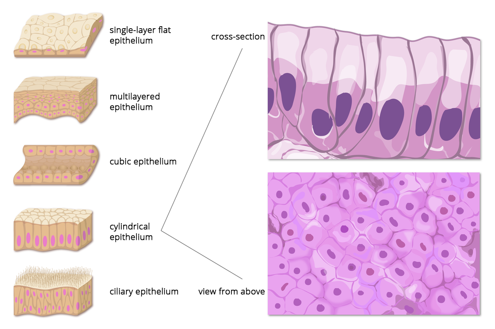
Type of epithelium (according to different classifications) | Examples of occurrence | Structure and properties | Tissue function |
stratified flat | epidermis, oral cavity | stratified, with tightly adjacent cells the outer layers of which can peel off | this flexible, relatively thick, dense shell protects the interior of the body (organ) |
simple flat | walls of blood vessels, alveoli | flattened cells, multi‑walled, close‑fitting | forms a thin layer that allows for the diffusion of respiratory gases and water, movement of nutrients, metabolites; in case of cornea – light penetration |
glandular | glands, gastrointestinal tract | typical cylindrical cells with a large number of Golgi apparatuses | produces digestive enzymes, tears, mucus, tallow, sweat |
ciliary | reproductive organs, trachea and bronchi, intestine | cylindrical cells have cilia on the surface facing the interior of the organ | cilia move the germ cells in the female reproductive system, transport impurities and mucus in the airways, increase the intestinal absorptive area |
Indicating adaptations of the structure of the epithelium of a frog's skin for the performance of its proper function.
permanent slide of epithelial tissue,
microscopic imaging equipment.
Students observe the frog's epithelial tissue through a microscope.
Recognise cell structures visible under a microscope, describe the shape of cells, recognise and identify cells that produce mucus.
Recall and list the functions of the frog’s skin.
Indicate the features of the tissue structure which are adaptations to performed functions.
Draw 2‑3 neighbouring cells and describe them. Remember that the drawing should be done in pencil and based on the image observed under the microscope as well as reflect the shape and proportions of the cells.
The frog skin epithelium is simple (single‑layer), which facilitates gas exchange (cutaneous respiration); it produces mucus that moistens the skin's surface, facilitating the diffusion of gases; its cells are compact and protect against the penetration of microorganisms.
Muscle tissues
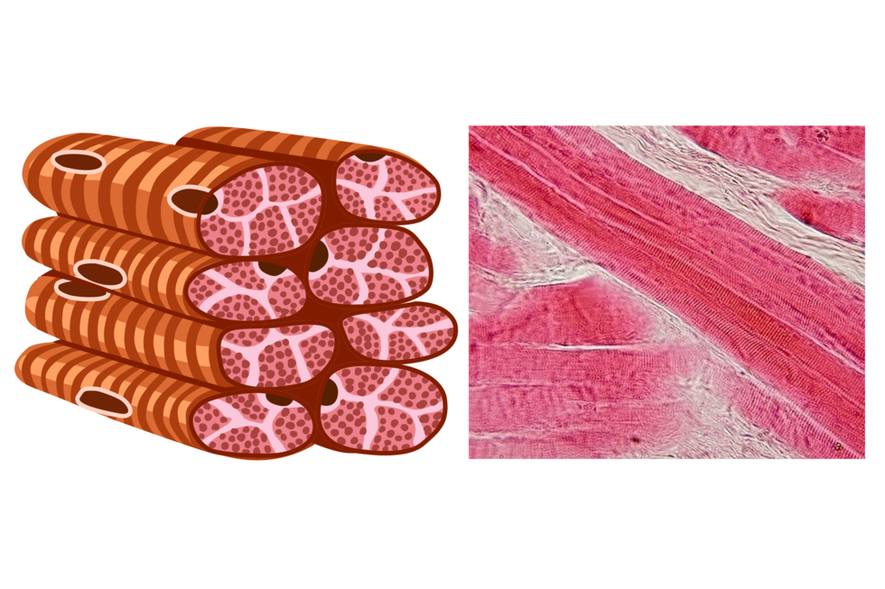
Due to the structure and location in the body, we distinguish three types of muscle tissue: striated skeletal muscle tissueskeletal muscle tissue cardiac muscle tissuecardiac muscle tissue and smooth muscle tissuesmooth muscle tissue. Muscle tissue is capable of performing contractions. Only the work of skeletal muscles is dependent on our will, thanks to which it is possible to perform intentional and conscious movements and to train the muscles themselves, taking care of their efficiency. The remaining muscles work constantly beyond our control at a rate appropriate to the level of metabolism. Smooth muscles are responsible, among others, for contractions of walls of the digestive tract during movement of the digestive tract contents and contractions of the walls of the blood vessels that cause changes in blood pressure. The muscles which the heart is made of shrink rhythmically, pumping blood into the arteries.
Muscle tissue cells are elongated. Their contractions are made possible by myofibrilsmyofibrils, built from proteins actinactin and myosinmyosin. The actin and myosin fibres cooperating with each other are shifting in relation to each other, which causes shortening of the cells and, as a result, muscle contraction.

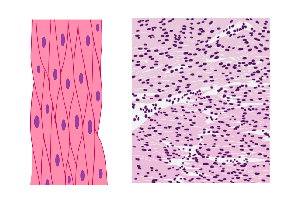
Features | Smooth muscle tissue | Striated skeletal muscle tissue | Cardiac muscle tissue |
Location | the walls of the gastrointestinal tract, blood vessels, reproductive organs, excretory ducts | locomotor system muscles, facial muscles, tongue, diaphragm | heart wall |
Shape of cells | spindle‑like, sharply ended | cylindrical, enlongated | cylindrical, enlongated, branched |
The number of nuclei in the cell | 1 | many | 1 |
Location of nuclei | central | peripheral | central |
Contraction regulation | independent of will | dependent on will | independent of will |
Contraction speed | low | very high | medium |
Function | internal organ walls movement, with the exception of the heart | maintaining body posture, body movement | heart work |
In comparison to other types of muscle tissues, cardiac muscle tissue has the highest number of mitochondria. It is also strongly supplied with blood. Taking into account the given structure features, explain what is the adaptation of this tissue to rhythmic contractions – from 6th week of embryonic life to the end of life.
Select the true sentences.
- Smooth muscles occur in stomach walls.
- The work of smooth muscles is dependant on our will.
- Striated muscles move eyelids.
- Muscles contain contractile proteins, actin and myosin.
- Muscle cells do not contain mitochondria.
- The epithelia line the body only from the outside.
- Interior of the oral cavity is covered by epithelial tissue.
- Epithelial tissue cells adhere closely to each other.
- Epithelial tissue has a strengthening function.
Organize the elements that build your body. Place the carbon atom at the very bottom.
- muscle cell
- muscular system
- locomotor system
- the human body
- arm muscle
- carbon atom
- fat molecule
Conclusion
The human body has got a hierarchical structure, which can be illustrated as follows: cell – tissue – organ – organ system – organism.
Individual structures of the body cooperate with each other, performing vital functions.
There are 4 types of tissues in the human body: epithelial, muscular, nervous, combined.
The structure of the tissue shows adaptations to the function performed.
Epithelial tissue performs many functions: protective, secretory, sensual, reproductive.
Muscle tissue can contract, which is why it is responsible for the movement function.
How was this lesson? Did you like it? Finish selected sentences.

Keywords
muscle tissue, epithelial tissue, organ
Glossary
aktyna – białko występujące w cytoplazmie komórek; umożliwia ruch i zmianę kształtu komórki; w mięśniach wspólnie z miozyną odpowiada za ich skurcz
miozyna – białko występujące w cytoplazmie komórek eukariotycznych; w mięśniach wspólnie z aktyną odpowiada za ich skurcz
miofibryle - włókienka kurczliwe; zbudowane głównie z białek: aktyny i miozyny.
tkanka – zespół komórek o podobnej budowie, funkcji i pochodzeniu
tkanka mięśniowa gładka – w jej budowie występują wrzecionowate komórki, przez nieregularne ułożenie włókien nie posiada prążkowana; działa niezależnie od naszej woli; jest odporna na zmęczenie; wstępuje w ścianach narządów, np. w żołądku.
tkanka mięśniowa poprzecznie prążkowana – składa się silnie wydłużonych, walcowatych komórek - włókien mięśniowych; układ miofibryli wykazuje poprzeczne prążkowanie; skurcze tej tanki są zależne od naszej woli; występuje w mięśniach szceletowych np. bicepsie.
tkanka mięśniowa poprzecznie prążkowana typu sercowego – zbudowana z wydłużonych, cylindrycznych komórek. Ułożenie włókienek kurczliwych wykazuje poprzeczne prązkowanie. Występuje w sercu, jej skurcze są nieżależne od naszej woli.