Eye - the organ of sight
organism is able to receive information and react to them;
in your brain there are sensory centres, which receive information form the receptors, as well as association areas, where the received information is interpreted;
neural impulse travels from the receptor through the sensory neuron, neural centre, efferent neuron to the effector.
to explain how the build of the eye is connected with its functions;
to describe, how visual sensations are created;
to explain, what different types of eye defects are and how we can correct them;
to explain selected types of eye illnesses, including their causes and symptoms;
to plan an experiment that proves the influence of light on the iris.
Sclera, choroid and retina
An eyeball has a spherical shape. Its outer layer is composed of three membranes. ScleraSclera surrounds the eyeball and protects it from injuries. It also gives it its shape. At the back part of the eyeball it is thick and opaque, whereas in the front of the eyeball, where we call it corneacornea, it is thin and transmits sun rays.
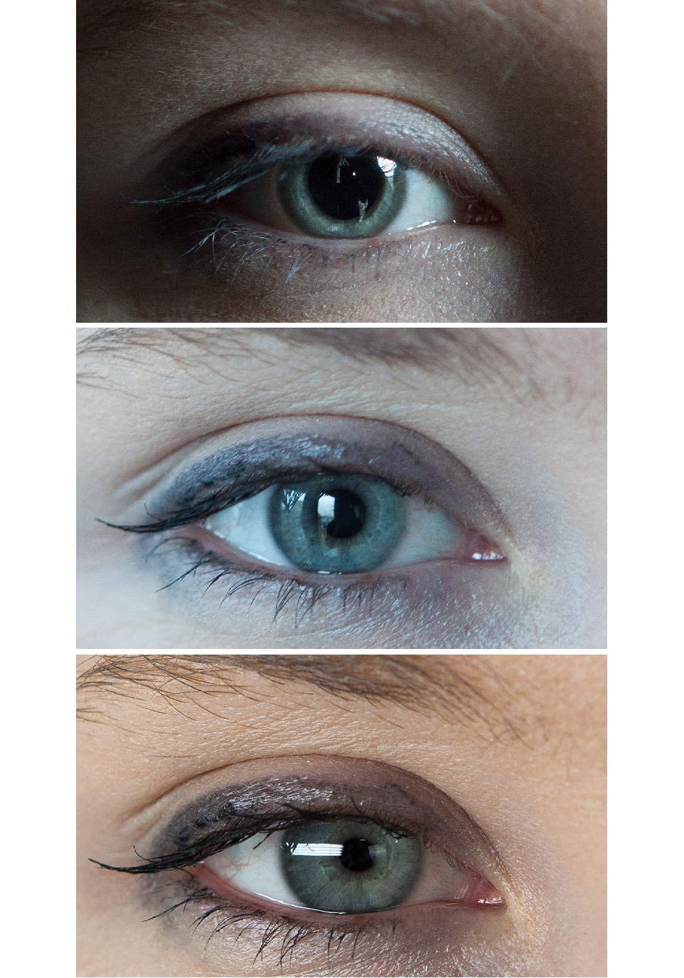
Under the sclera there is the choroidchoroid, which has blood vessels, it surrounds the eye and provides it with oxygen and products of metabolism. In the front part of the eyeball, the choroid converts into the iris, which has the shape of a ring. The opening in that ring is the pupil. In the iris there are smooth muscles, thanks to which it can change the diameter of the pupil and regulate the amount of light transmitted into our eye. Adapting to the intensity of light is reflex. The colour of the iris depends on the amount of the pigment – melanin.
The most internal membrane of the eyeball is the retinaretina. It has 2 different types of sensory cells that are sensitive to lightsensory cells that are sensitive to light – cone cells and rod cells. Rod cellsRod cells are sensitive to the intensity of the light – thanks to them our brain differentiates between different shades of gray, but not colours. Cone cellsCone cells ensure chromatic vision and are active only when the light is bright. This is the reason why we don’t see colours well in semidarkness. Each cone cell is responsible for catching light of different colour (wave lenght). On the retina there is 125 million rod cells and 6 million cone cells, whereas rod cells are found on the outside part of the retina and cone cells are found at the rear part of the eyeball. Its highest concentration (250 thousand per mmIndeks górny 22) is found in the point called macula luteamacula lutea. It is the area of the sharpest vision. On the retina there is also a place that has no sensory cells. It is the blind spotblind spot, from which departs the optic nerve that transfers neural impulses to the encephalon.
Confirm the presence of the blind spot on the retina.
you,
pen,
white sheet of paper.
In the middle of the paper, horizontally in one line draw a star (on the left) and a circle (on the right). Both should be around 1 cm in diameter.
Close your left eye and using your right eye look at the star.
Gradually move the picture away from you. Can you see the circle all the time?
Assess the distance of the paper at which the circle becomes invisible.
Repeat this observation for the other eye. This time, focus your eye on the circle and observe how the star disappears.
If the circle disappeared from the field of vision, it means that the light rays coming from the circle reached the part of the retina that is not sensitive to light.
How images are created
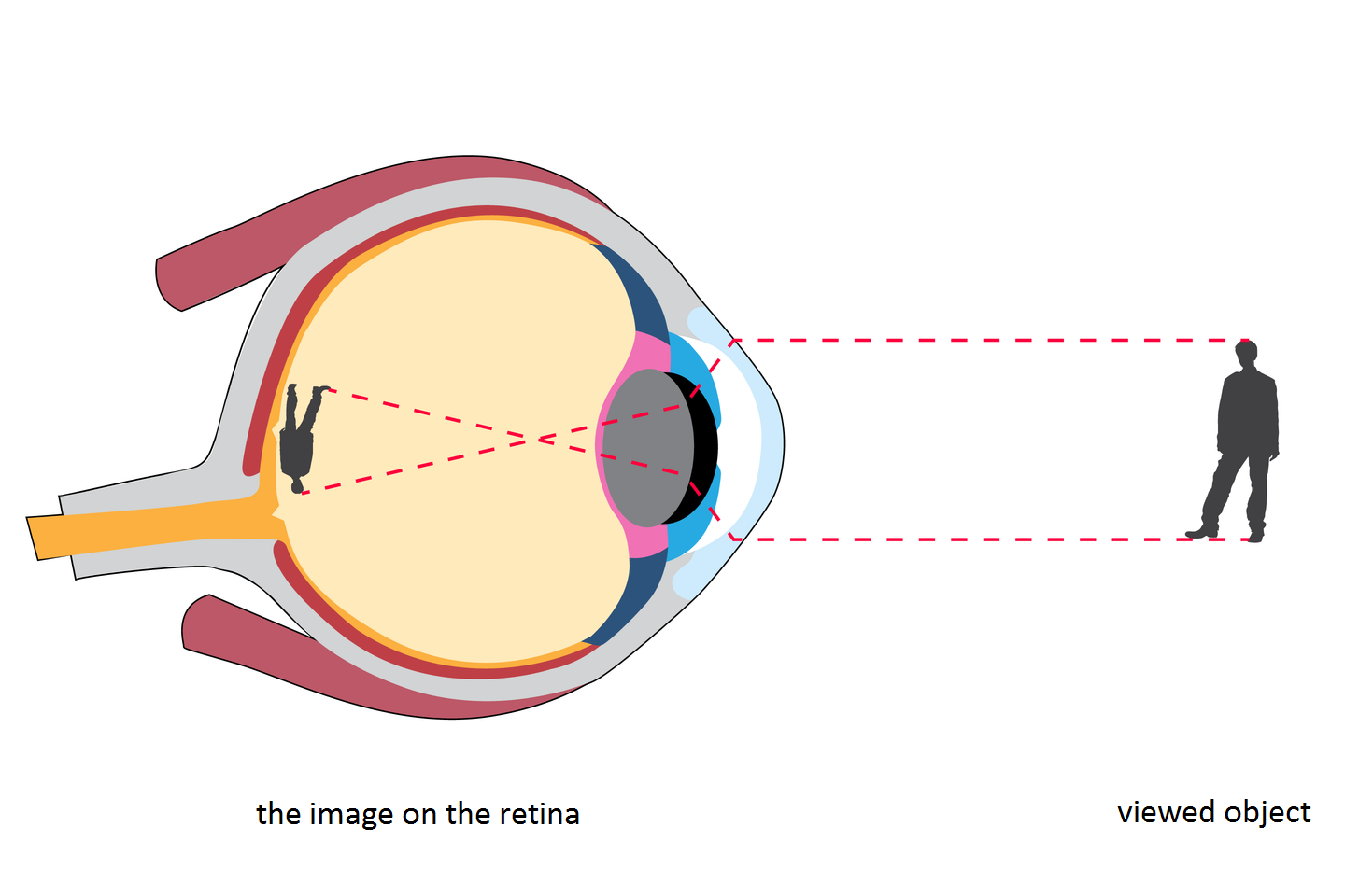
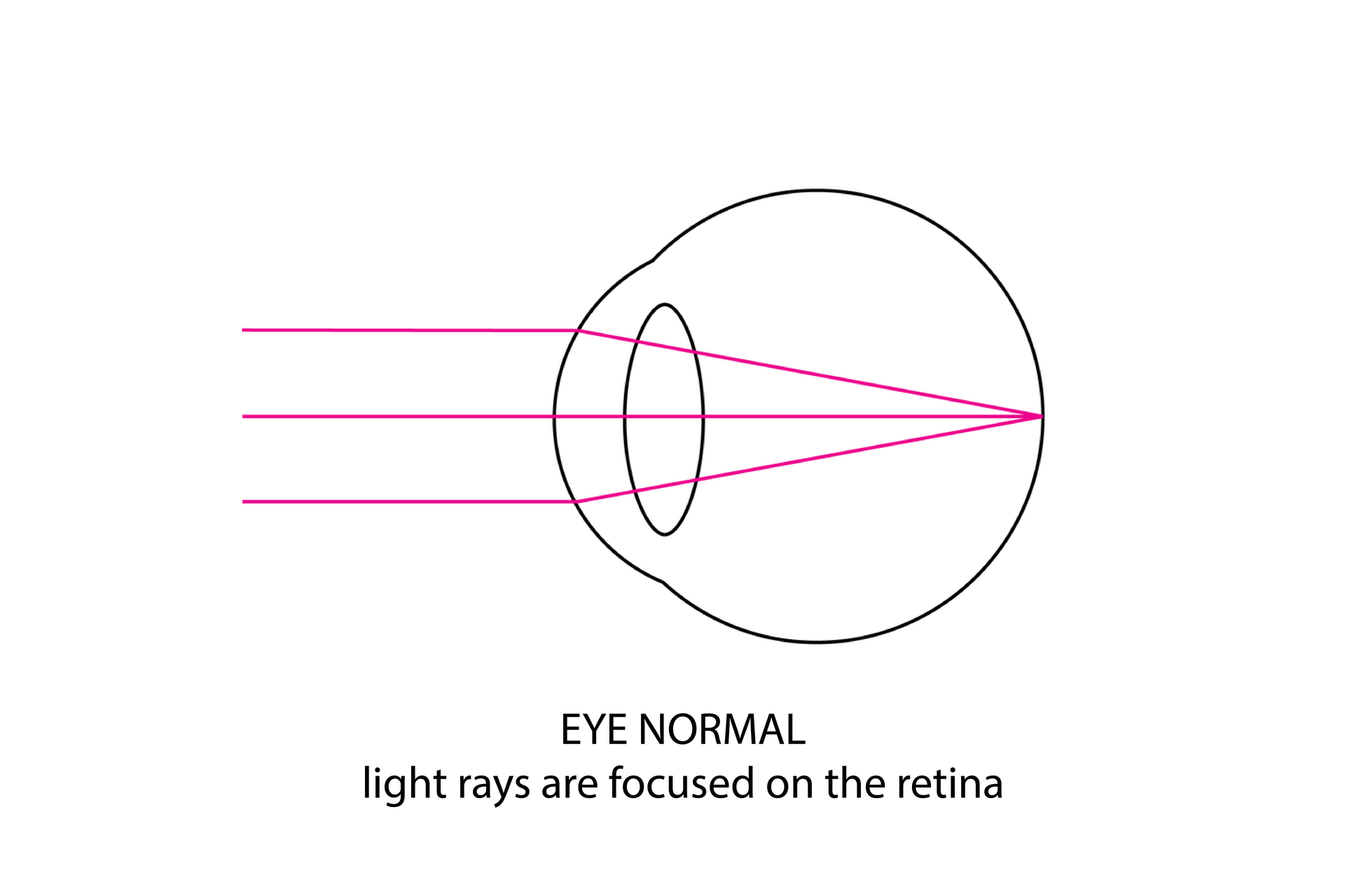
After passing through the cornea, the light reaches the pupil behind which is the lens. It is transparent and its shape can change. Sun rays go through the lens and then through the vitreous humour (vitreous body) that lies directly behind it and that consists in 98% of water. Next, they reach the surface of the retina. On the way, they become refracted by the cornea and by the lens, thanks to which the sharp image is created on the retina.
Light stimulates receptor cells on the retina. Each one of them has photosensitive pigment which, under the influence of the light energy, decomposes and causes the creation of a neural impulse. The impulse from the receptor cells travels to the optic nerve and then, through the optic nerve, to the optic cortex in the brain. There, neural impulses are translated and interpreted (recognized), thanks to which we learn how our surroundings look like.
Eye accommodation to seeing in different conditions
The most sensitive part of the retina is the macula lutea which consists mainly of cone cells. Image which is created there is sharp, clear and colorful. In complete darkness the cone cells become less sensitive, and the rod cells, situated at some distance from the macula lutea, become stimulated. The image created thanks to the rod cells is less sharp and gray.
Eyes adjust (adapt) to the amounts of light. After leaving a dark place into the light, and from a light room into the dark room, our eyes may be blinded for a short time, but then they begin to send information to our brain. Strong light can damage our eyesight, which is why we should not look directly into the sun without protective lenses. Strong light is a stimulus that causes us to squint and causes our pupil to narrow.
Tracking objects that are approaching or departing is possible thanks to the shape of the lens (curvature). It allows you to adjust the visual acuity. When we look at the image from up close, the lens becomes more convex, whereas when you are looking at the object from a distance, the lens becomes flatter. This phenomenon is called accommodationaccommodation of the eye.
Seeing on both eyes allows us to judge the distance. Eyeballs are directed towards the front, they are at distance of about 6 cm from each other. Because of that, they receive 2 slightly different images. Optic nerves transfer them to the brain and there, in the optic centre, both information are analyzed and superimposed on each other. This is how the spatial (three dimensional) image is created.
Organ of sight - illnesses and defects
Eyes deliver about 70% of information from the surroundings. Every day many external factors influence them, which is why they require special care and protection. Hygiene of the organ of sight should include those actions, which allow the eyes to function correctly. These are, among other things:
protecting the eyes from too intense light by using sunglasses with UV filter;
protecting your eyes from having a contact with chemical substances in water (e.g. in a swimming pool) by using swimming glasses;
remembering about correct moistening of the eyeball, by drinking around 2.5 l of liquids every day and maintaining adequate humidity in rooms, especially during heating season;
caring for correct lighting of your work place and positioning the computer screen at a distance not shorter than 50 cm from the eyes, and, when you work longer using a computer – using glasses with anti‑glare lenses;
limiting time we spend in front of the TV or computer;
regular eye checks.
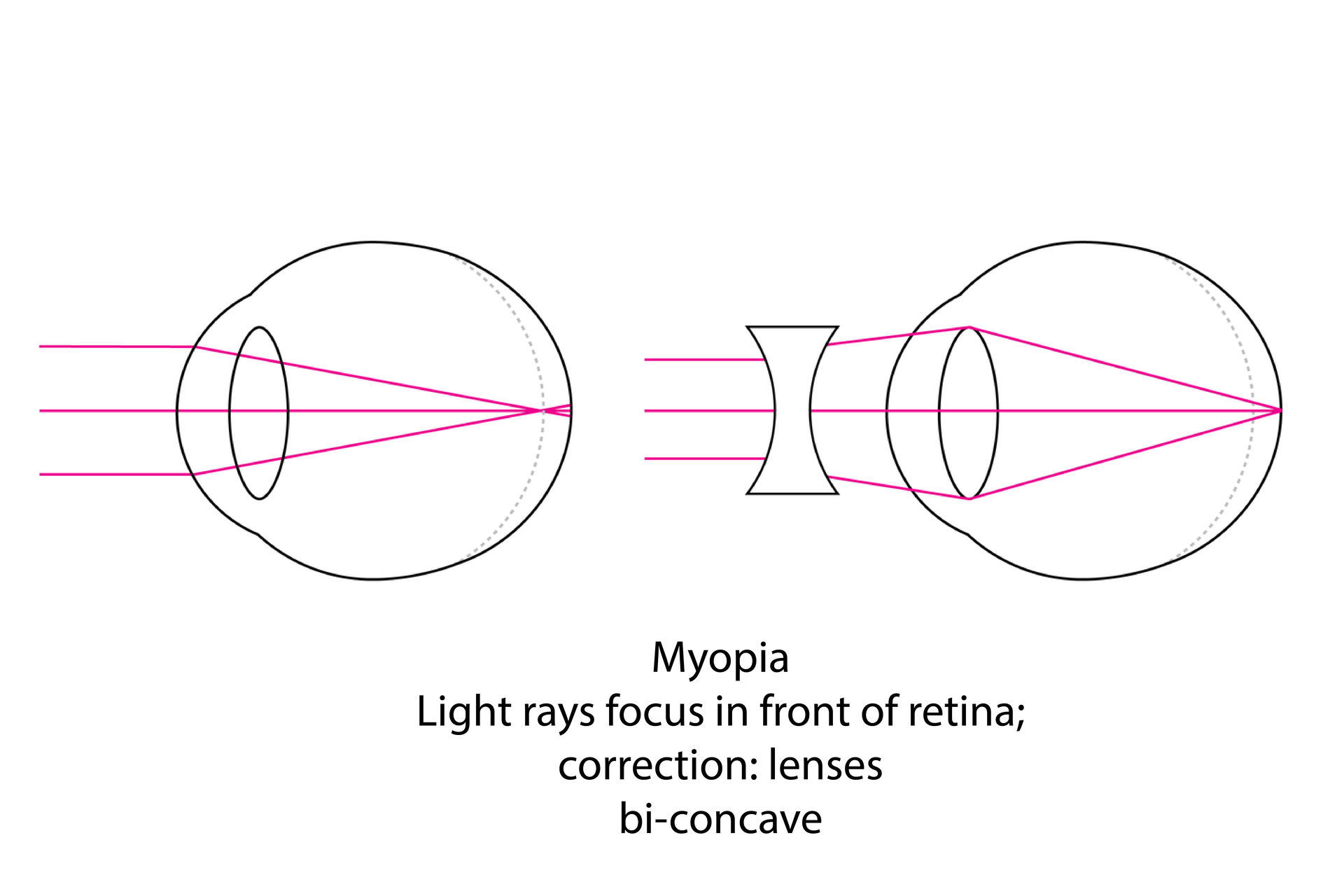
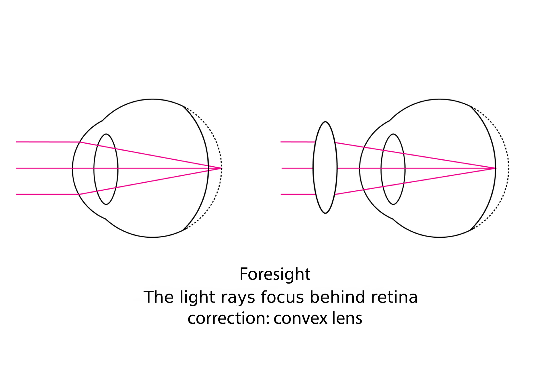
Wrong shape of the lens or the eyeball is the cause of myopia or hypermetropia, and wrong curvature of the elements that refract the light leads to astigmatism. Eyesight defects can be corrected using adequate lenses.
The cause of myopiamyopia can be the incorrect, too bulgy shape of the lens or too elongated eyeball. As a result, the light is focused before the retina, which causes the person with such defect to see clearly only the objects that are at a short distance from her eyes. In order to correct this defect, we use dispersive lenses, marked with the minus sign. In the case of hypermetropiahypermetropia, the eyeball is shortened or the lens is too flat, which causes the light to focus behind the retina. In order to correct this defect, we use focusing lenses, marked with the plus sign. The ability of the eye to see is expressed in diopters.
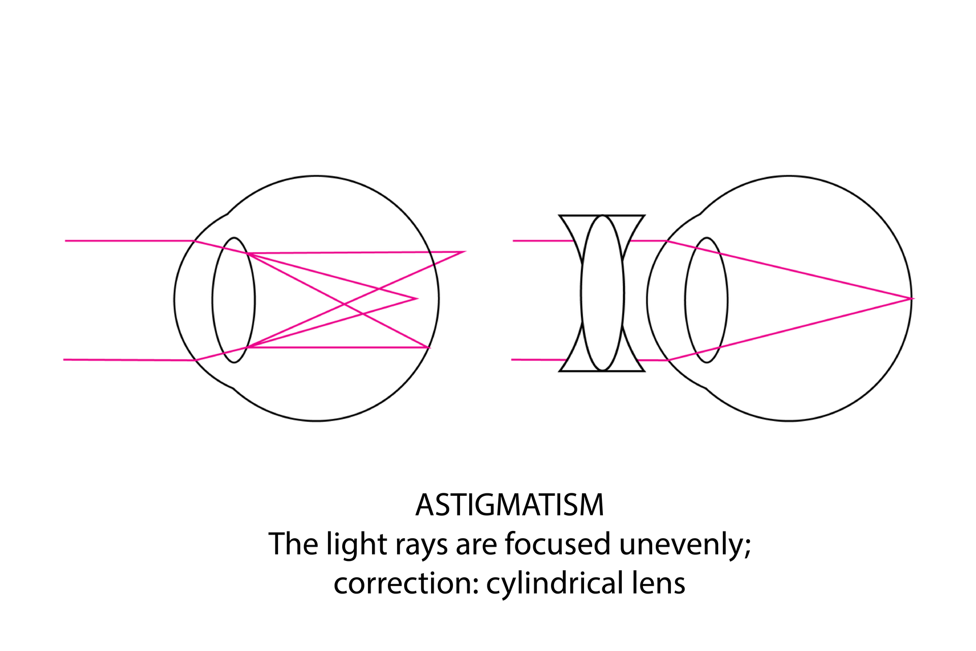
The irregular curvature of the cornea or the lens focuses the light in different points of the retina or outside the retina, which causes the image to be blurry. This type of defect is called astigmatismastigmatism. In order to correct it, we use cylindrical lenses.
Working with a computer requires focusing your eyes on a screen that is at fixed distance, which, in consequence, may lead to weakening of the muscles that are responsible for changing the shape of the lens and problems with eyes’ ability to accommodate. That is why it is recommended to make short breaks while working, during which we should look at something far away. Looking at a computer screen lowers the frequency of blinking, which leads to bad moistening of the eyeball surface. In this situation, you feel your eyes burning, or you have the impression you have sand in your eyes. If this discomfort continues, it can lead to cornea infections.
Arrange the elements and present the flow of information from the cornea to the cerebral cortex. Place the cornea on top.
- retina
- cornea
- optic centre in the brain
- vitreous body
- pupil
- optic nerve
- lens
Summary
Organ of sight consists of the eyeball and the protective and moving apparatus.
Eyeball is covered by three membranes: sclera, choroid and retina.
On the retina there are photoreceptor cells - cone cells and rod cells.
Macula lutea is the place of sharpest vision.
Light passes through the cornea, lens, vitreous humour (vitreous body) and reaches the retina.
The image created on the retina is smaller and inverted.
The phenomenon of accommodation allows us to see sharply objects that are close and far.
Problems with sight can be caused by eye diseases, genetic conditions or result from wrong construction and shape of the elements of the eye.
Keywords
eye, retina, pupil, lens
Glossary
akomodacja – zjawisko polegające na zmianie kształtu soczewki, zapewniające ostrość widzenia przedmiotów z bliska i daleka
astygmatyzm – wada wzroku, w której nieregularny kształt krzywizny rogówki lub soczewki powoduje, że promienie świetlne skupiają się w wielu różnych punktach
czopki – komórki zmysłowe wrażliwe na barwy światła: czerwoną, zieloną, niebieską; działają tylko przy dobrym oświetleniu
dalekowzroczność – wada wzroku, w której zbyt krótka gałka oczna lub zbyt płaska soczewka powodują, że promienie świetne skupiają się za siatkówką
fotoreceptory – u kregowców komórki reagujące na światło; znajdują się w siatkówce oka; należą do nich pręciki i czopki
krótkowzroczność – wada wzroku, w której zbyt długa gałka oczna lub zbyt wypukła soczewka powodują, że promienie świetne skupiają się przed siatkówką
naczyniówka – cienka błona leżąca między twardówką a siatkówką, zaopatrzona w naczynia krwionośne; dostarcza do siatkówki substancje odżywcze i tlen
plamka ślepa – miejsce na siatkówce pozbawione fotoreceptorów, z którego wychodzi nerw wzrokowy
plamka żółta – miejsce na siatkówce o największym skupisku czopków; stanowi punkt najostrzejszego widzenia
pręciki – komórki zmysłowe wrażliwe na natężenie światła i ruch; są aktywne nawet przy bardzo słabym oświetleniu
rogówka – przezroczysta przednia część twardówki; błona w przedniej części gałki ocznej, okrywająca tęczówkę i źrenicę
siatkówka – błona wewnętrzna gałki ocznej, zawierająca komórki światłoczułe – czopki i pręciki
twardówka – błona okrywająca od zewnątrz gałkę oczną; chroni ją przed uszkodzeniami mechanicznymi oraz nadaje jej kształt